3D Dental Imaging in Katy
State-of-the-art 3D dental imaging in Katy, TX at Orange Tree Oral Surgery. Precise imaging technology ensures accurate planning and treatment.
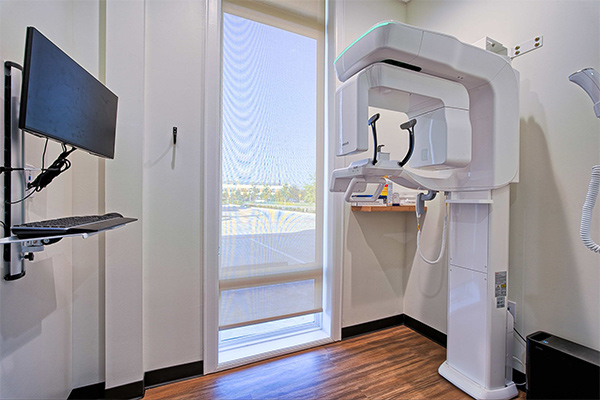
What is 3D Imaging?
Our practice uses Cone Beam Computed Tomography (CBCT) as our 3D imaging of choice. CBCT is a specialized imaging technique used in dentistry to produce 3D images of the teeth, jaws, jaw joints, and surrounding structures. It provides detailed and comprehensive information that is valuable for diagnosis, treatment planning, and evaluation of various conditions.
How Does Cone Beam Computed Tomography (CBCT) Differ From Traditional Dental X-rays?
Unlike traditional dental X-rays, which produce 2D images, CBCT generates a 3D volume of the scanned area. This allows for better visualization of anatomical structures, more accurate measurements, and improved assessment of spatial relationships. Additionally, CBCT images can be viewed from different angles and planes, providing a more comprehensive understanding of complex anatomy.
Why Do I Need a CBCT Scan?
CBCT scans are commonly used in oral surgery for various purposes, including:
- Implant planning
- Assessing impacted teeth
- Diagnosing temporomandibular joint (TMJ) disorders
- Evaluating the jawbone for pathology or abnormalities
How Much Radiation Does CBCT Use?
CBCT uses multiple techniques to lower radiation used. Typically CBCT uses about 0.01 to 0.1mSv. This amount of radiation is comparable to getting a single chest X-ray and about 1/50 to 1/100 the radiation from a medical-grade CT scan.